
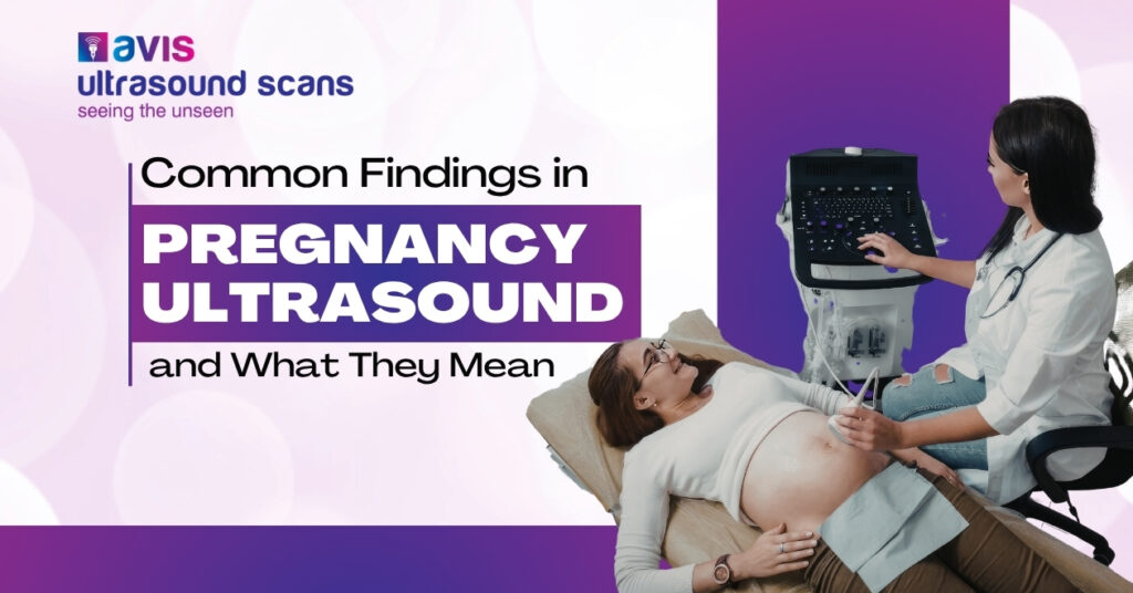
An early pregnancy ultrasound is often one of the most reassuring moments for expecting parents. It confirms the pregnancy, helps estimate gestational age, and checks whether early development is progressing normally. At the same time, the scan may show findings that raise questions or cause concern if their meaning isn’t clearly explained.
At Avis Ultrasound Scanning Centre, early pregnancy ultrasound scans in Hyderabad are performed to provide clarity, reassurance, and timely medical guidance. This blog explains the common findings seen in an early pregnancy ultrasound and what they usually mean.
What is an Early Pregnancy Ultrasound?
An early pregnancy ultrasound is typically done between 5 and 10 weeks of pregnancy. Depending on how early the scan is required, it may be performed as:
- Transvaginal ultrasound (common in very early weeks)
- Abdominal ultrasound (as pregnancy progresses)
The goal of this scan is to confirm pregnancy location, assess early development, and rule out complications.
Gestational Sac
The gestational sac is usually the first structure seen on ultrasound, often visible around 4.5 to 5 weeks of pregnancy.
What it means:
- Confirms that pregnancy is present
- Confirms the pregnancy is inside the uterus
- Helps estimate early gestational age
A normally placed gestational sac is a reassuring sign. If no sac is seen when expected, doctors may recommend repeat scanning or blood tests.
Yolk Sac
The yolk sac appears shortly after the gestational sac, usually between 5 to 6 weeks.
What it means:
- Indicates a developing pregnancy
- Provides early nutrition to the embryo
- Helps assess pregnancy viability
A normal-sized yolk sac is reassuring. Abnormal size or shape may require closer monitoring, but does not always indicate a problem.
Fetal Pole
The fetal pole represents the earliest visible form of the embryo and is usually seen by 5.5 to 6 weeks.
What it means:
- Confirms embryonic development
- Allows accurate dating of pregnancy
- Used to measure crown–rump length (CRL)
If the fetal pole is not yet visible, it may simply be too early. Repeat ultrasound after a week often provides clarity.
Fetal Heartbeat
Detecting the fetal heartbeat is one of the most important milestones in early pregnancy. It is usually visible between 6 to 7 weeks.
What it means:
- Strong indicator of a viable pregnancy
- Helps assess early fetal well-being
- Heart rate provides information about development
A heartbeat that is slightly slower early on may normalize as pregnancy progresses. Absence of heartbeat very early does not always indicate loss — timing matters.
Crown–Rump Length (CRL)
CRL measures the length of the embryo from head to bottom and is the most accurate method for dating early pregnancy.
What it means:
- Determines gestational age
- Helps estimate expected delivery date
- Confirms growth is appropriate for weeks of pregnancy
CRL measurement is more reliable than last menstrual period dates, especially if cycles are irregular.
Subchorionic Hematoma
Sometimes, ultrasound shows a small subchorionic hematoma, which is a collection of blood near the pregnancy sac.
What it means:
- Common finding in early pregnancy
- Often resolves on its own
- May be associated with spotting or bleeding
Most subchorionic hematomas do not harm the pregnancy, but doctors may recommend rest and follow-up scans.
Multiple Gestational Sacs
Early ultrasound may reveal more than one gestational sac, indicating a twin or multiple pregnancy.
What it means:
- Confirms multiple pregnancy early
- Helps determine chorionicity (shared or separate placenta)
- Allows proper pregnancy monitoring from the start
At Avis Ultrasound Scanning Centre, with the best female radiologist in Nallagandla, early detection helps in planning proper care and monitoring your baby’s growth from the very beginning.
Corpus Luteum Cyst
A corpus luteum cyst is often seen on one ovary during early pregnancy.
What it means:
- Normal hormonal structure
- Supports pregnancy in early weeks
- Usually disappears on its own
This is a normal and expected finding and typically does not require treatment.
Empty Gestational Sac (Early Stage)
Sometimes, a gestational sac is seen without a yolk sac or fetal pole.
What it means:
- May simply indicate very early pregnancy
- Requires follow-up ultrasound
- Not immediately a cause for concern
Repeat scanning after 7–10 days usually clarifies the situation.
Why Follow-Up Ultrasounds Matter ?
Early pregnancy develops rapidly, and findings can change within days. Follow-up scans help:
- Confirm normal progression
- Monitor uncertain findings
- Provide reassurance
- Guide timely medical decisions
At Avis Ultrasound Scanning Centre, we provide trusted pregnancy ultrasound scans in Nallagandla with careful attention to detail, accurate interpretation, and clear communication so you feel informed and reassured at every step.
Schedule Your Early Pregnancy Ultrasound
Early pregnancy ultrasound provides valuable insight into the very first stages of life. Most findings are normal variations of early development, and understanding what they mean helps reduce unnecessary anxiety. Clear explanations, proper timing, and expert evaluation make all the difference in early pregnancy care.
If you have been advised an early pregnancy ultrasound or need reassurance during the first weeks of pregnancy, accurate imaging is essential.
Call +91 88774 69999 to book your scan at Avis Ultrasound Scanning Centre, Nallagandla.
Early clarity brings peace of mind and we’re here to support you every step of the way.